Laptop Ultrasound
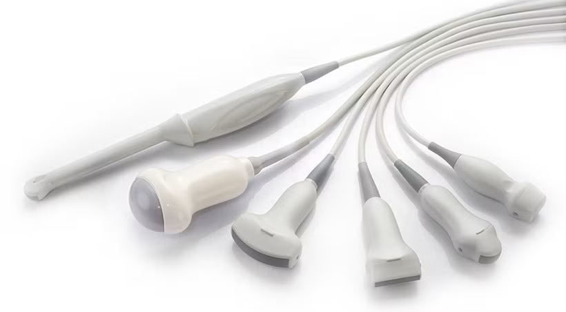
Probes
Supports various types of probes such as linear array, convex array, phased array, micro-convex, volume, intracavity and biplane, etc.
Laptop Ultrasound Specifications
1. Support multiple operating modes such as B, 2B, 4B, CF, PW, M, CW, CE, NE, PDI, DPDI, TDI, 3D/4D, EFOV, AM, etc. and their combination modes.
2. Supports various types of probes such as linear array, convex array, phased array, micro-convex, volume, intracavity and biplane, etc.
3. 8-segment TGC and LGC are adjustable.
4. Provide multi-department measurement and calculation inspection reports.
5. Ultra-wide frequency band and up to 256 elements.
TFM(Total Focusing Method)
Total focusing method (TFM) is an ultrasonic array technology used to comprehensively focus each point in a region of interest. TFM is a processing method of data collected by synthetic focusing using FMC, which involves reconstruction within a region of interest (a frame of pixels) to focus on many points forming a grid.
1. Ultra-wideband uniform beam emission
Total focusing imaging breaks the beam shape limitation of traditional emission “fixed-point focusing” and performs “intelligent dynamic beam control” on the entire area. Obtain consistent high resolution across the entire field of tissue without frame rate loss.
2. Stronger and more uniform energy
Traditional ultrasound energy is mainly concentrated near the selected focus, and the energy will decay rapidly away from the focus. Total focusing imaging technology breaks through this limitation through real-time correlated energy change emission methods, and makes the energy distribution in the entire field uniform.
3. Excellent penetration
The resolution of traditional ultrasound images is limited by the focus position and the propagation speed of sound waves in tissues. Total focusing imaging is based on the RF metadata platform. It performs holographic correction and acoustic beam optimization processing on the massive real-time changing
Total focusing imaging
whole-field RF metadata, further achieving extraordinary detail resolution and excellent penetration at high frequency and high resolution, thereby obtaining high quality images.
Total focusing imaging breaks through the technical limitations of traditional ultrasound imaging platforms, solves the “out-of-focus blur” problem of traditional ultrasound imaging, and brings clinicians better image quality, more accurate diagnostic tools, and more intelligent clinical applications experience.
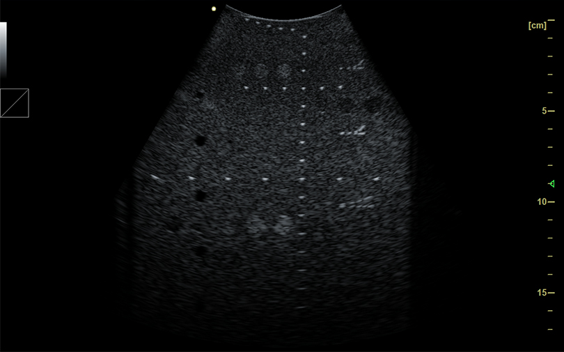
L18M probe imaging
Ultra-fast Imaging
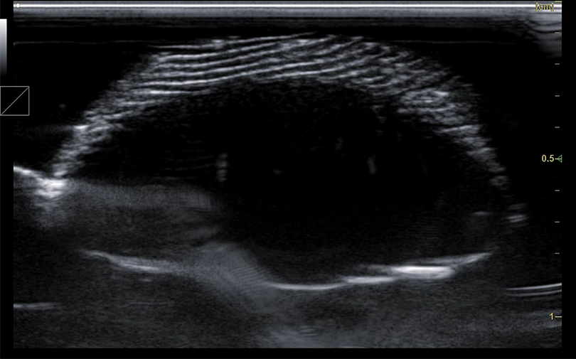
Ultrafast ultrasound imaging breaks through the frame rate limitations of traditional ultrasound imaging and can capture higher-resolution, higher-definition images at a frame rate 100 times faster than traditional imaging. This technology will bring advancements in prevention, diagnosis, and treatment monitoring.
CMUT Technology
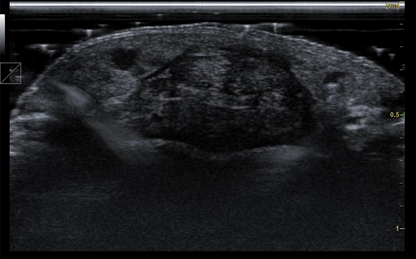
Using CMUT(Capacitive Micromachined Ultrasonic Transducer) semiconductor ultrasound technology, the spatial resolution of the image is increased to micron-level high-definition imaging, providing clear two-dimensional images for clinicians to observe and diagnose the epidermis, dermis, subcutaneous tissue and skin appendages. Rich and sensitive color Doppler blood flow imaging satisfies the judgment of subcutaneous blood flow and skin tumor status.
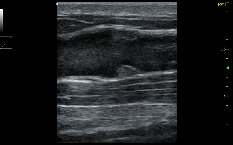
L30M probe imaging
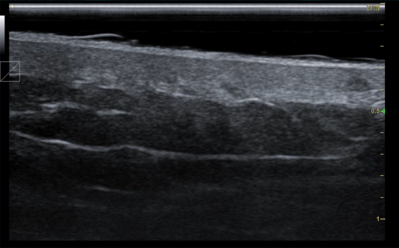
Below are some excellent clinical images.
Ultra-fast ultrasound imaging
CMUT ultrasound technology imaging
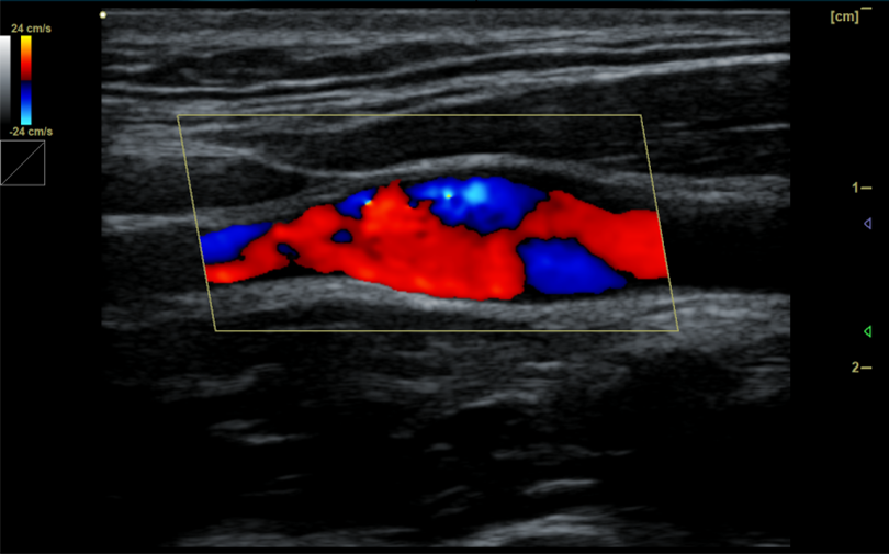
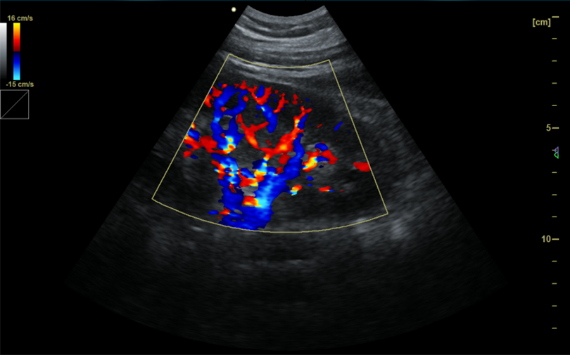
Carotid bulb blood flow
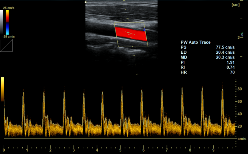
PW auto trace
Abdominal aorta B image
Abdominal aorta blood flow
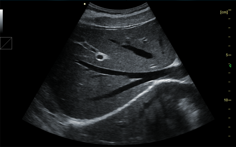
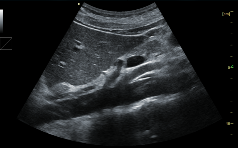
Liver B image
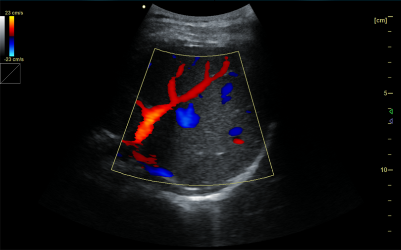
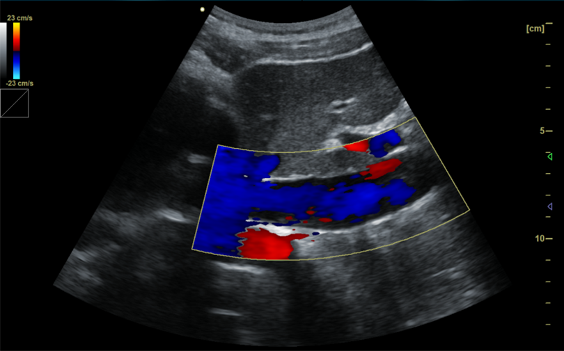
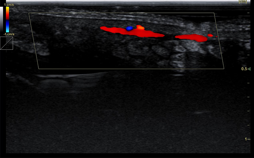
Liver blood flow
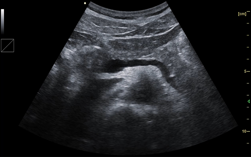
Sarcoskeleton
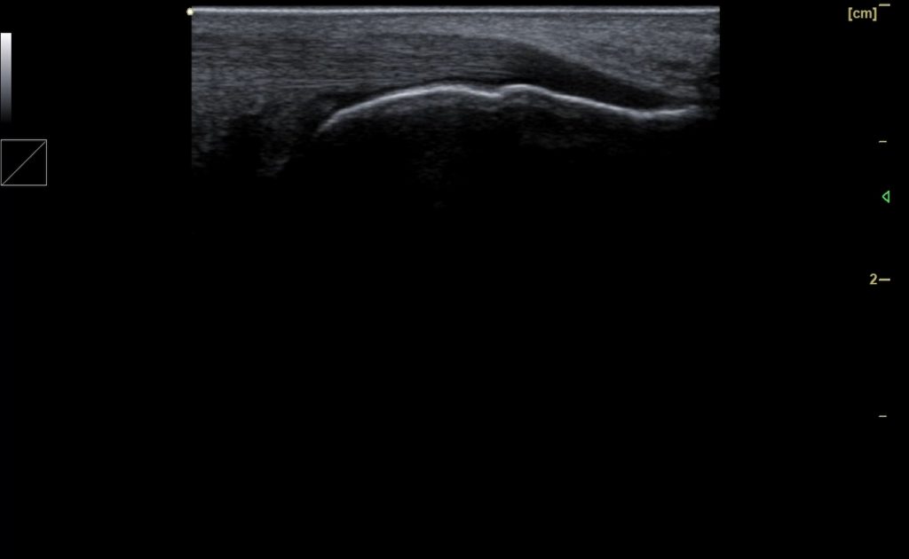
Fingerprint
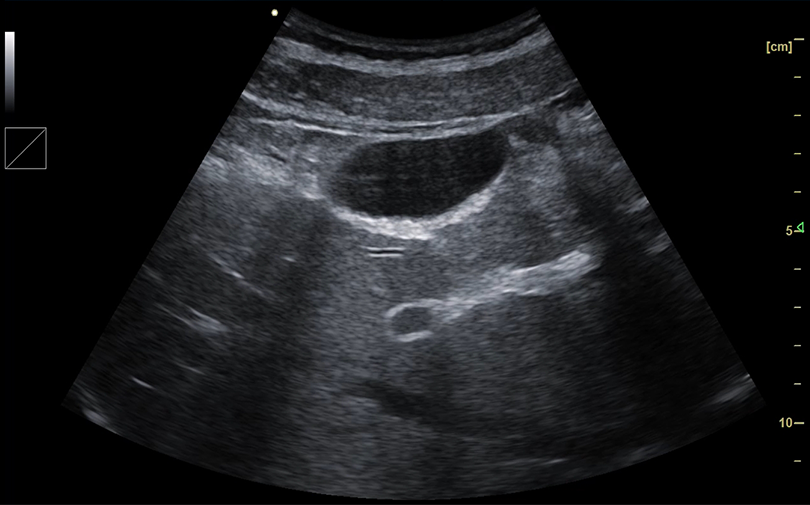
Gall bladder
Spleen
Nail bed blood flow
Bursae bursae calcaneus retrocalcaneus
Kindey blood flow
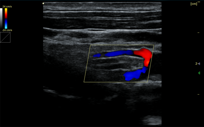
Vertebral artery blood flow
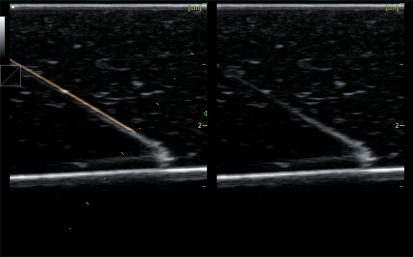
Needle enhancement
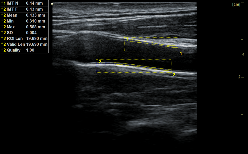
IMT automatic measurement and calculation
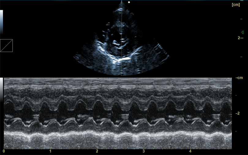
Small animal heart M image
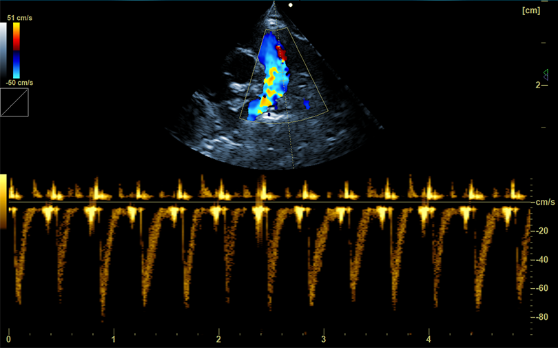
Small animal heart Doppler image
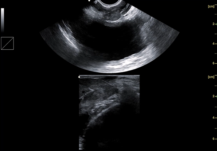
Biplane B image
F.A.Q.
Lorem ipsum dolor sit amet.
Lorem ipsum dolor sit amet, consectetur adipiscing elit. Ut elit tellus, luctus nec ullamcorper mattis, pulvinar dapibus leo.
Lorem ipsum dolor sit amet, consectetur adipiscing elit. Ut elit tellus, luctus nec ullamcorper mattis, pulvinar dapibus leo.
Lorem ipsum dolor sit amet, consectetur adipiscing elit. Ut elit tellus, luctus nec ullamcorper mattis, pulvinar dapibus leo.
Lorem ipsum dolor sit amet, consectetur adipiscing elit. Ut elit tellus, luctus nec ullamcorper mattis, pulvinar dapibus leo.
Lorem ipsum dolor sit amet, consectetur adipiscing elit. Ut elit tellus, luctus nec ullamcorper mattis, pulvinar dapibus leo.