Portable Ultrasound
Portable Ultrasound Specifications
1. Support multiple operating modes such as B, CF, PW, M, NE, PDI, DPDI and their combination modes.
2. Supports various types of probes such as linear array, convex array and phased array.
3. Applicable to Android, ios(only for wireless probes) and windows operating systems.
4. Provides simple measurements and calculations.
5. Support DICOM function..
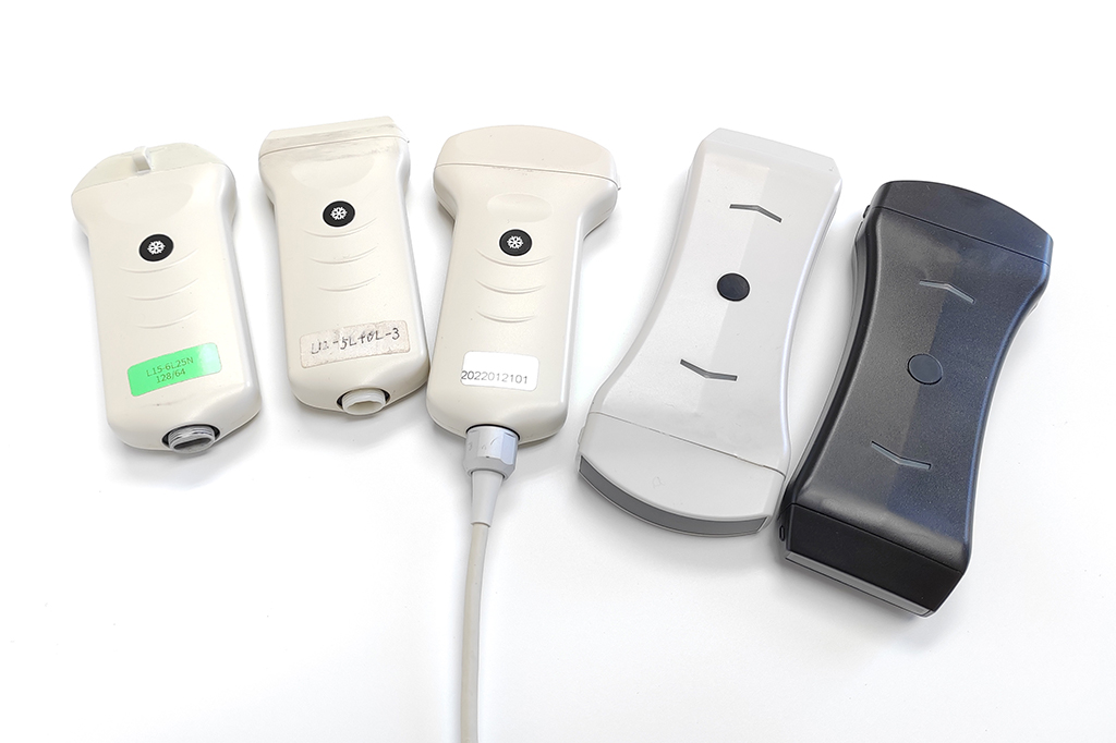
Probes
Supports various types of probes such as linear array, convex array, phased array, including wired and wireless probes.
Composite B image
Real-time Composite Imaging
Spatial composite technology can effectively reduce speckle noise, thereby making uniform tissue images smoother and more delicate. It can also significantly improve the signal-to-noise ratio and contrast of the image, which is beneficial to clinicians’ diagnosis. In addition, scanning with different deflection angles can obtain information at different angles and detect interfaces in different directions, after spatial compounding, the image information is richer and the interface continuity is better. Another important application of spatial composite technology is puncture needle display enhancement. Through spatial composite deflection scanning, the incident sound beam is made as perpendicular to the surface of the puncture needle as possible, thereby obtaining a strong puncture needle surface image.
Wireless Dual Probe
Portable ultrasonic double-head series probes can be combined with two probes at the same time to achieve the functions of two probes at the same time. The probe switching function is implemented in the software, which avoids problems such as too many physical button functions that are difficult to operate.
Double probe switching
Probe presets
Probe Presets
There are default, carotid artery, thyroid, finger, liver, kidney and other presets. Selecting the corresponding preset operation will reduce the user’s examination time and improve examination efficiency.
Below are some excellent clinical images.
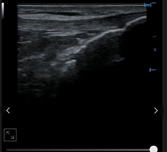
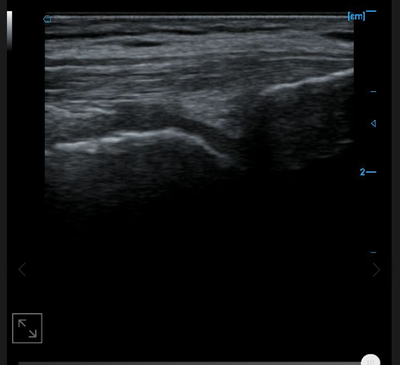
wireless images
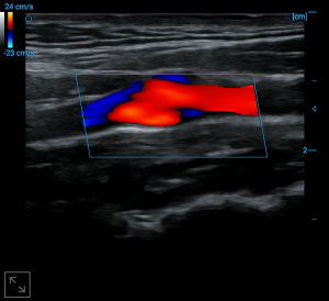
Carotid bulb blood flow
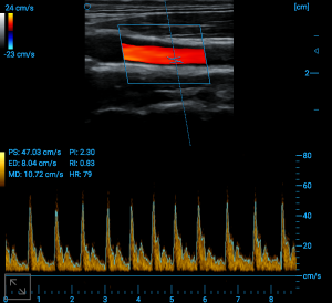
PW auto trace
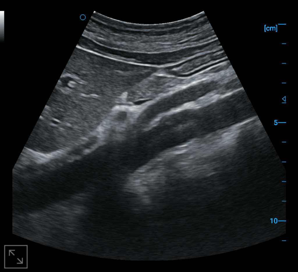
Abdominal aorta B image
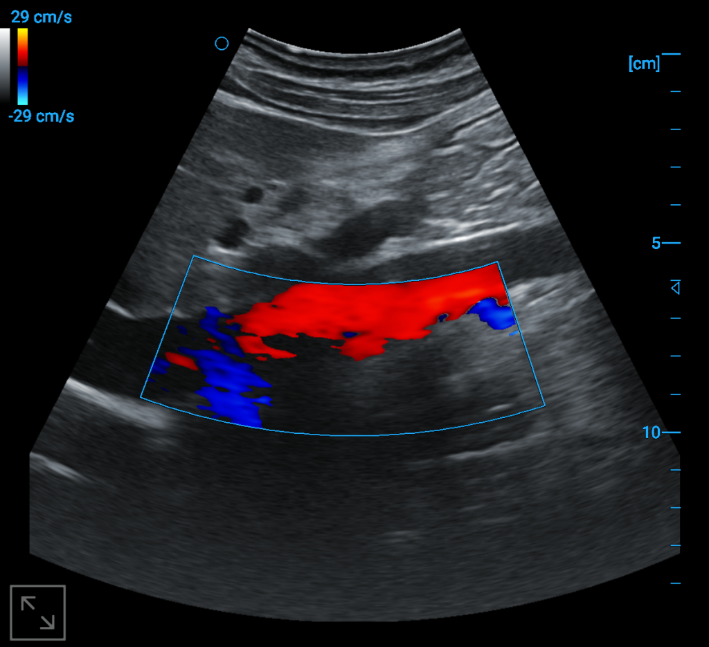
Abdominal aorta blood flow
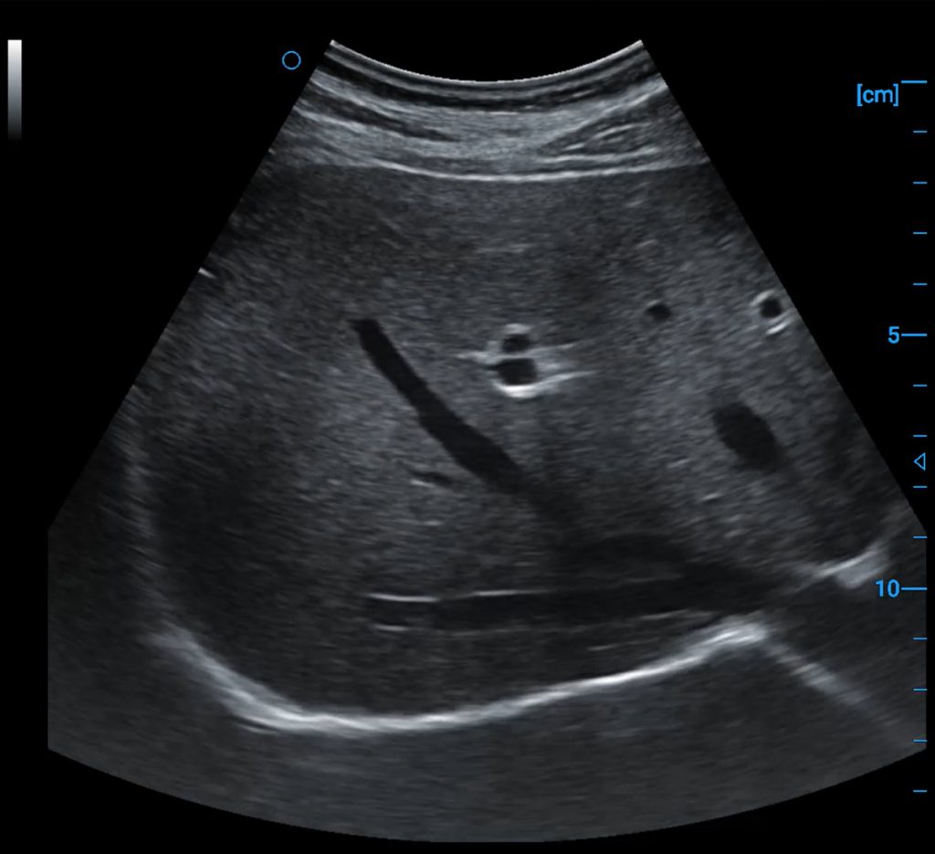
Liver B image
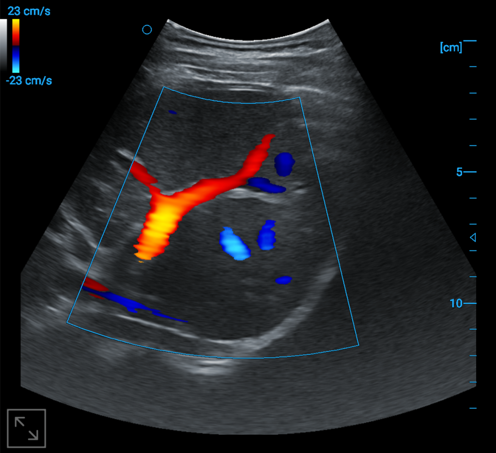
Liver blood flow
wireless images
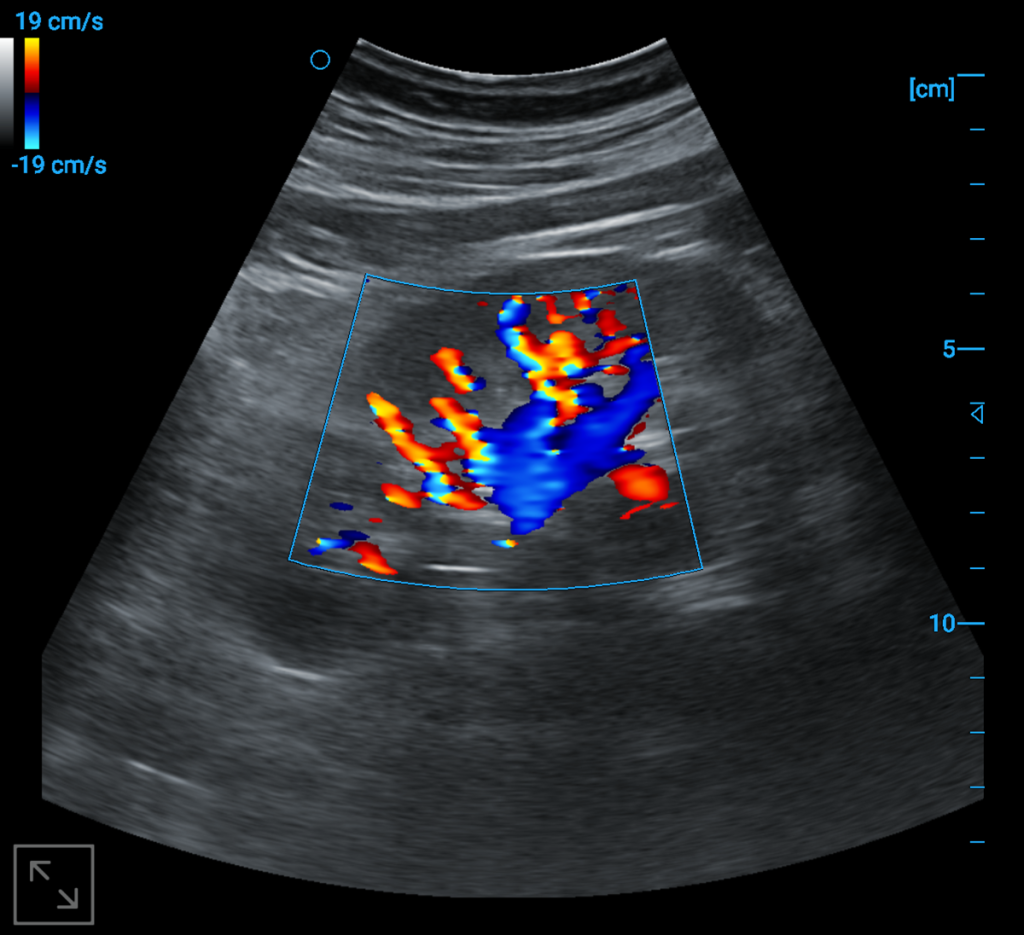
Kindey blood flow
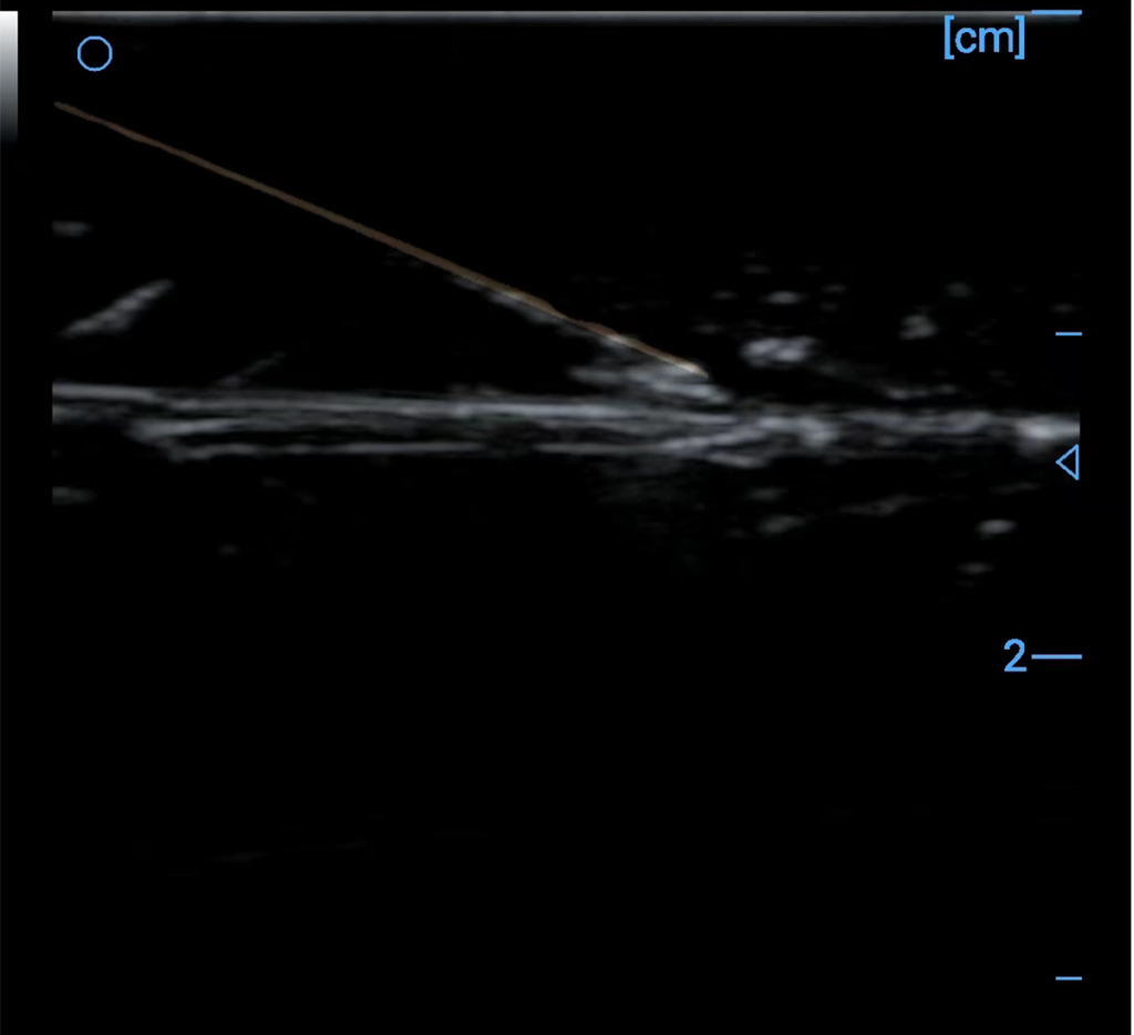
Needle enhancement
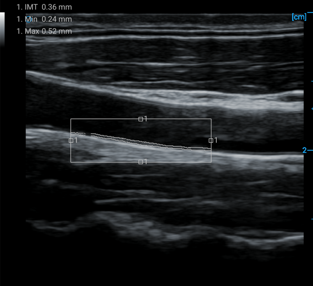
IMT automatic measurement and calculation
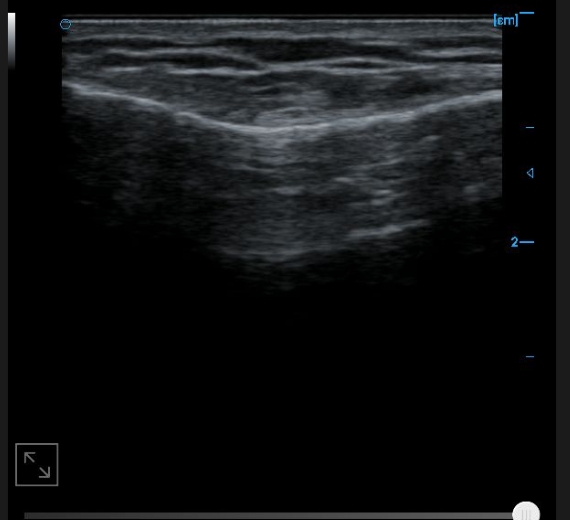
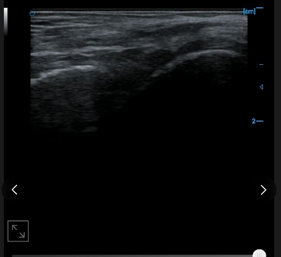
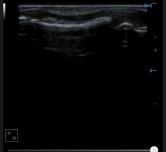
wired images
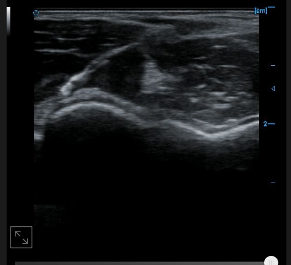
Patellar tendon B image
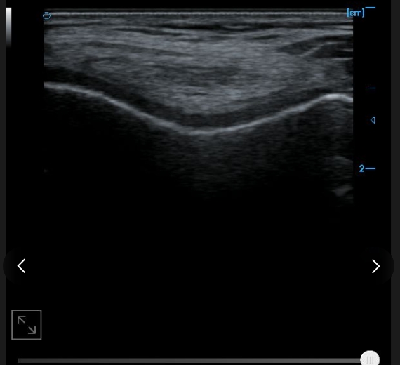
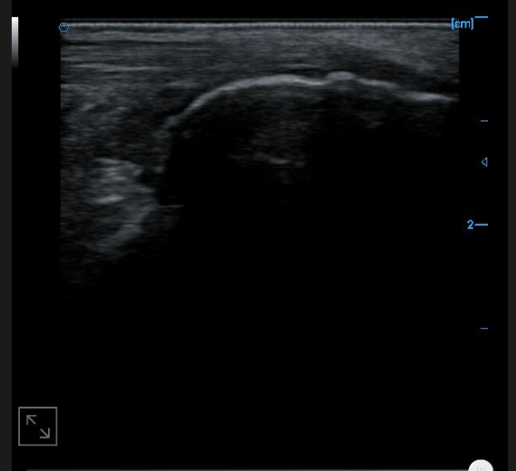
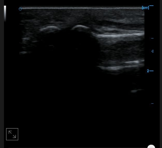
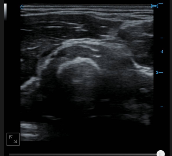
B-image of quadriceps tendon
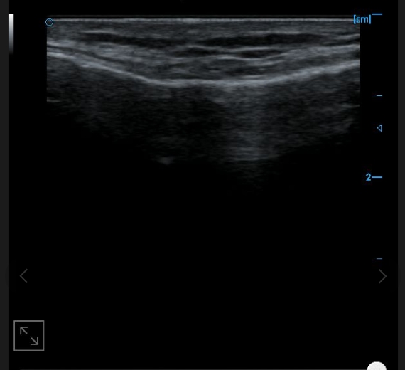
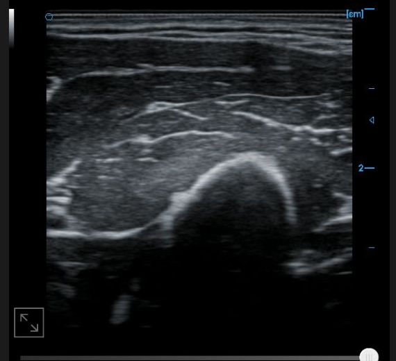
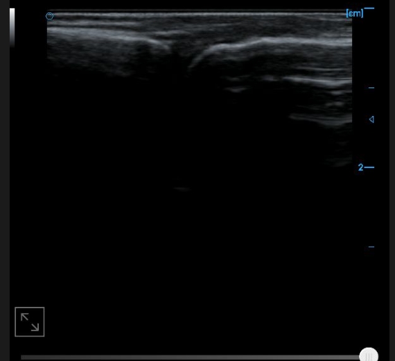
Knee collateral ligament B image
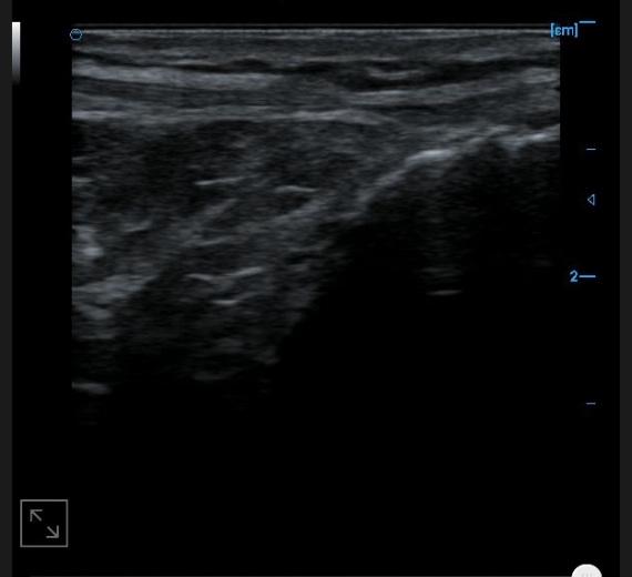
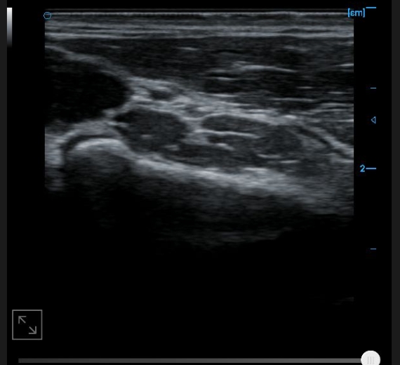
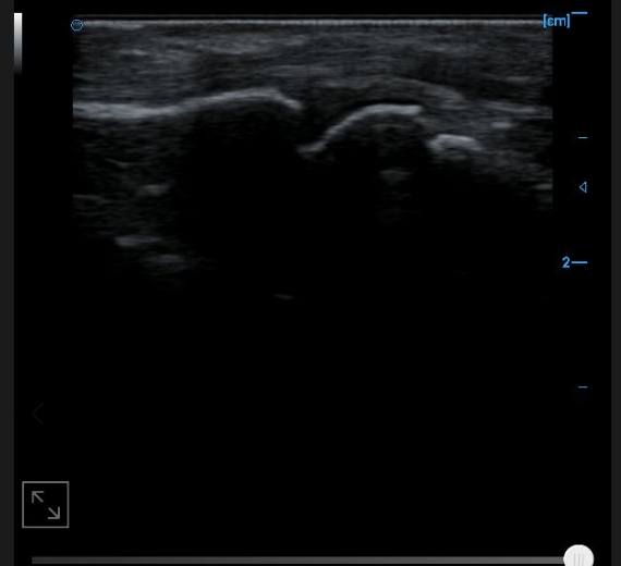
B-image of femoral trochlear cartilage
Internal testing supports images with B
External testing supports images with B
Posterior tibial tendon B image
Posterior calcaneal bursa B image
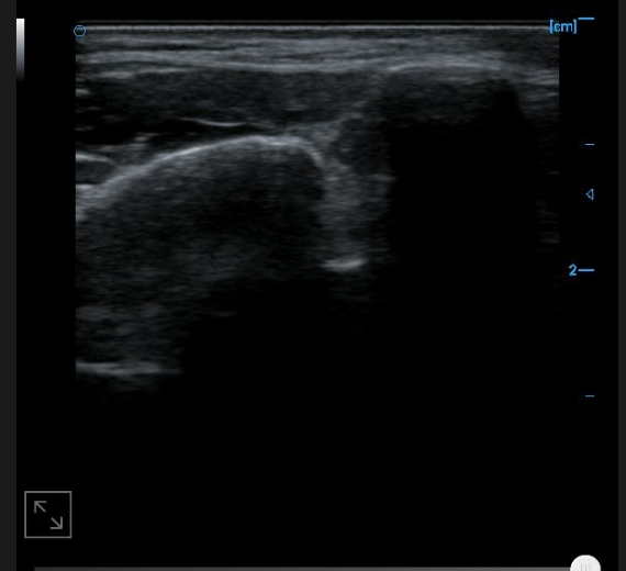
Image of anterior tibiofibular ligament B
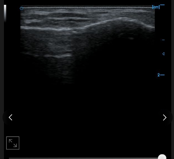
Image of anterior talofibular ligament B
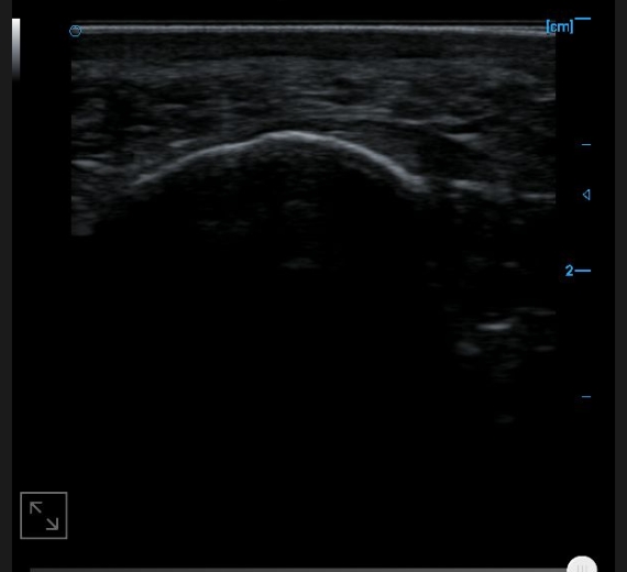
Image of plantar aponeurosis B
B image of toe and flexor digitorum tendon
B-image of the anterior cross-section of the upper arm
Cross sectional B-image of the distal tendon of the biceps brachii muscle
B-axis imaging of the distal tendon of the biceps brachii muscle
B-image of upper forearm cross-section
Finger extensor tendon B image
A1 Trochlear and joint recess B image
Finger flexor tendon B image
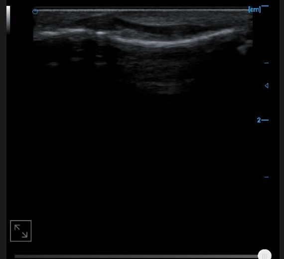
Image of flexor digitorum profundus tendon B
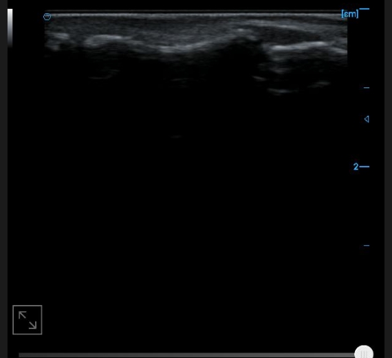
Image of lateral collateral ligament B
F.A.Q.
Lorem ipsum dolor sit amet.
Lorem ipsum dolor sit amet, consectetur adipiscing elit. Ut elit tellus, luctus nec ullamcorper mattis, pulvinar dapibus leo.
Lorem ipsum dolor sit amet, consectetur adipiscing elit. Ut elit tellus, luctus nec ullamcorper mattis, pulvinar dapibus leo.
Lorem ipsum dolor sit amet, consectetur adipiscing elit. Ut elit tellus, luctus nec ullamcorper mattis, pulvinar dapibus leo.
Lorem ipsum dolor sit amet, consectetur adipiscing elit. Ut elit tellus, luctus nec ullamcorper mattis, pulvinar dapibus leo.
Lorem ipsum dolor sit amet, consectetur adipiscing elit. Ut elit tellus, luctus nec ullamcorper mattis, pulvinar dapibus leo.